This is a learning note related to the modilarity in neuroimaging. It contains the basic physics principles behind them and difference between them.
Modilarity introduction
Three types of medical imaging modalities are available to us:
- External: X-Ray Radiographs (X-Ray), X-Ray Computed tomography (CT). Shoot the X-ray go and through the body,
- Internal: Single Photon Emission Tomography (SPECT), Positron Emission Tomography (PET). Inject something radioactive into body, then it goes wherever we wanted. The electron shine out of the body and be measured.
- External and Internal: Magetic Resonance Imaging (MRI). Use some radio-waves from the outside to stimulate sth on the inside. Then look at the radio waves that the hydrogen atoms in the body emit back.
Basic physics
- Radiation: emission of energy as electromagnetic waves, aka photons, or as moving subatomic particles, aka alpha particles, electrons or positrons
- Ionizing radiation: radiation with sufficient energy to cause ionization in the medium through which it passes
- Ionisation: process in which an atom or a molecule acquires a negative or positive charge by gaining or losing electrons to form ions
- Radioactive decay: Unstable (that is, radioactive) nuclei can become more stable after emitting particles and energy. This process is called Radioactive decay. These emitted particles or energy (the latter one emitted by electromagnetic waves) are collectively referred to as radiation.
- Types of Radioactive decay:
- $\alpha$ - alpha particle = helium nucleus, 4-8 MeV (big size), very low penetration, piece of paper stops it
- $\beta$ - electron or positron emission, few MeV, also low penetration -> PET
- $\gamma$ - by-product of radioactive decay, 100-200 keV, higher penetration -> SPECT
X-Ray and CT

1. the difference between gamma and x-rays
- basis of wavelength with radiation: between $10^{-9}$ to $10^{-11}$ defined as X-rays and $10^{-11}$ to $10^{-13} $defined as gamma rays.
- radiation source: X-rays are emitted by electrons, while gamma rays are emitted by the atomic nucleus
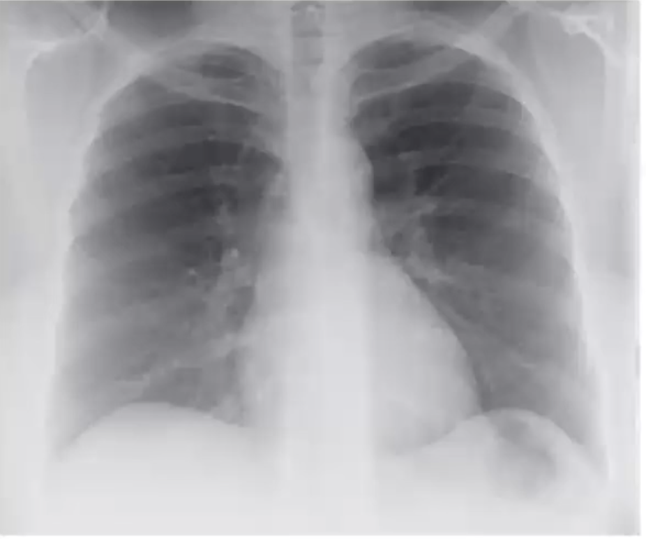
2. how an X-ray image is formed
It is more like taking a photo. X-Ray shoot on the body, it go thorough the body and depends on the different density between different part of the body to get a grey image. The lighter part means some part with heigh density, while darker part presents some part with low density. Contrast depends on the density, resolution depends on the number of particles shot on the body, signal to noise ration (SNR) depends on how long the scan takes,
3. formula for the radon transform
Radon transform is an integral trasform which is considered as sum of intensities of pixels in each direction.
Given objective function $f(x,y)$, change the coordinate system with $xcos\theta + sin\theta = p$ and $-xsin\theta + ycos\theta = q$
Radon thranform is: \(P(p, \theta) = R{f(x,y)} = \int_Lf(x,y)dl = \int_{-\infty}^{\infty}f(pcos\theta-qsin\theta, psin\theta+qcos\theta)dq\)
4. schematic diagram describing backprojection
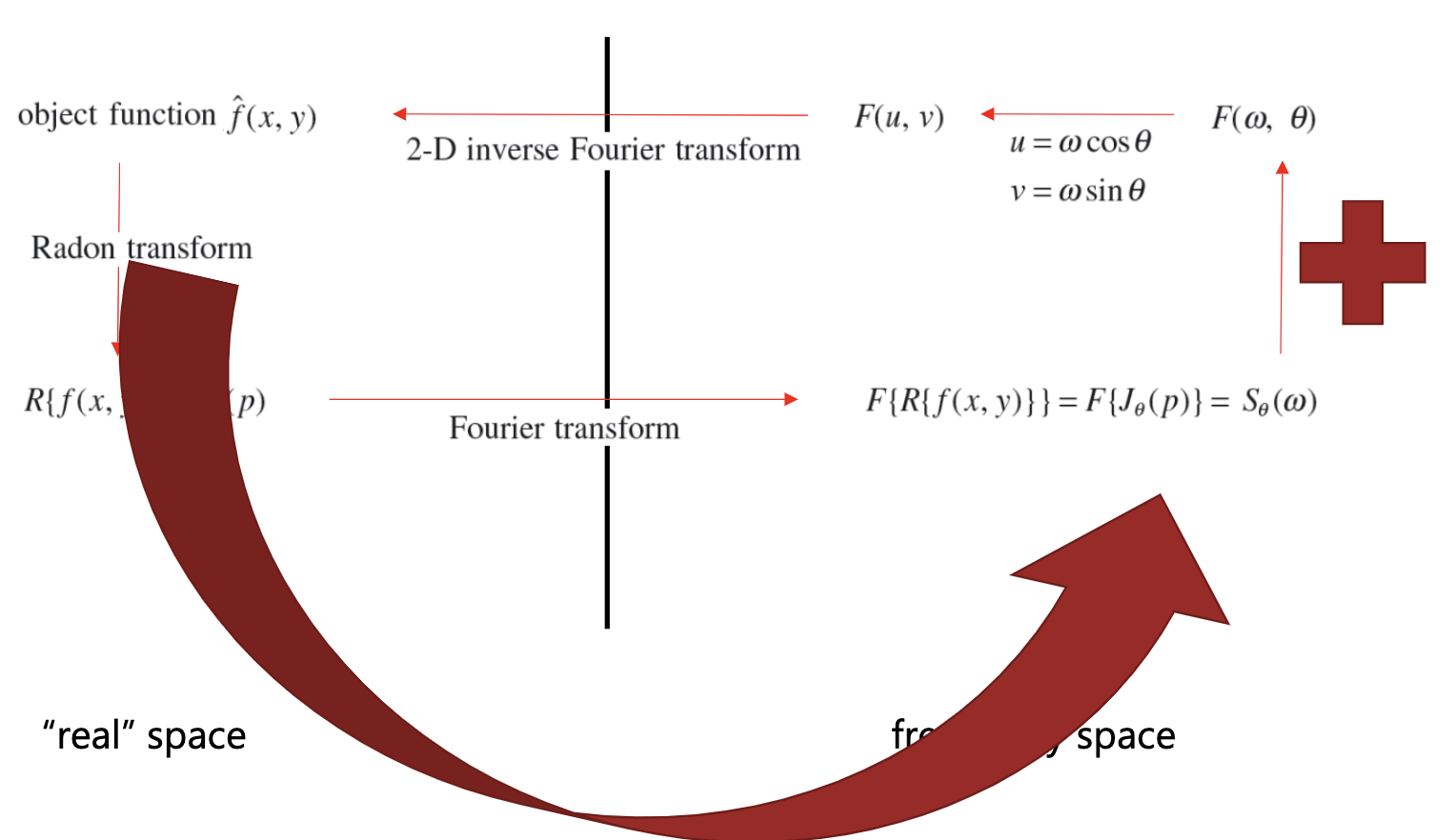
5. how filtered backprojection differs
Use filter to limit the spacial frequency that we want to see in image. e.g. filter out low frequencies to avoid blurring.
MRI
1. The difference between MRI and X-Ray or CT
- MRI combines external and internal sources, it uses an external mechanism (radio waves) to excite atomic nuclei that rotate in a magnetic field (resonance). When the atomic nuclei return to their previous energy level, radio waves are emitted (internal) that can be measured.
- provides high-resolution images with excellent soft -tissue contrast superior to X-ray & CT because of the underlying physics of the imaging process
- contrast in the MRI signal is based on the difference of hydrogen protons in the tissue and physiological structures
- can image anatomical structures as well as biochemical properties based on physiological function, including blood flow and oxygenation
- MRI is harmless as it does not use ionizing radiation, in contrast to ordinary X-ray or computer tomography
- BUT patients with magnetic metal in the body or a pacemaker cannot be examined with MRI due to the strong magnetic field, and patients with claustrophobia may have difficulties undergoing MRI.
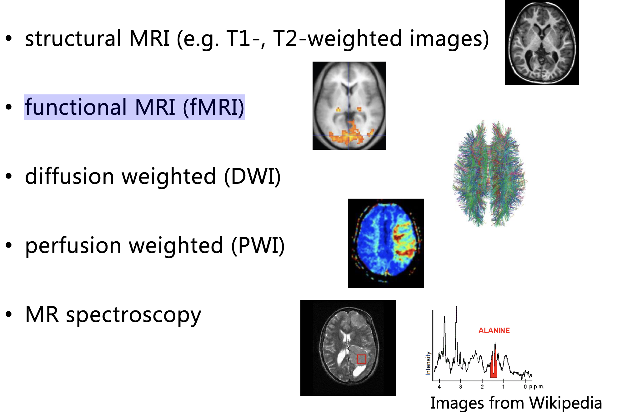
2. Different types of MRI images
-
structural MRI (e.g. T1-, T2-weighted images)
white part - white matter in the brain
grey part - cortical ribbon where all neural neuralons are located
black part - ventricles
-
functional MRI (fMRI)
reflects blood oxygenation levels. Some brain activity going on there
3. Basic Physics
MRI uses the nuclear magnetic resonance (NMR) property of the matter of the object
- Nuclear Magnetic Resonance (NMR): the nuclei spins in in a strong constant magnetic field. When an additional oscillating magnetic field is added, the nuclei is disturbed. Then remove the additional filed, we ca capture the energy released by the nuclei in the process of returning back to the original state. Finally we get NR signals which can help distinguish different tissues.
- When the object is placed into the MR scanner and this causes the nuclei to align with the magnetic field. The nuclei process about the field at similar frequencies, but at a random phase with respect to each other. This causes net longitudinal magnetization in the direction of field. So within a slice, a radio frequency (RF) pulse is used to align the phase and tip over the nuclei, which causes the longitudinal magnetization to decrease and establishes a new transversal magnetization. After the RF pulse is removed, the system seeks to return to eguilibrium. The transverse magnetization disappears, and the longitudinal magnetization grows back to its original size. During this process of returning to the equilibrium, a signal is created that can be used to construct the image.